Ultrasound is one of the important methods to help monitor fetal development throughout pregnancy. Currently, 2D and 4D ultrasound are both commonly used in obstetrics, each technique has its own advantages and complements each other during fetal monitoring.
2D ultrasound is a traditional method, creating black and white images according to cross-sections of the fetus. This technique helps doctors evaluate gestational age, fetal position, fetal heart, placenta, amniotic fluid, development index, and detect many structural and morphological abnormalities of the fetus.
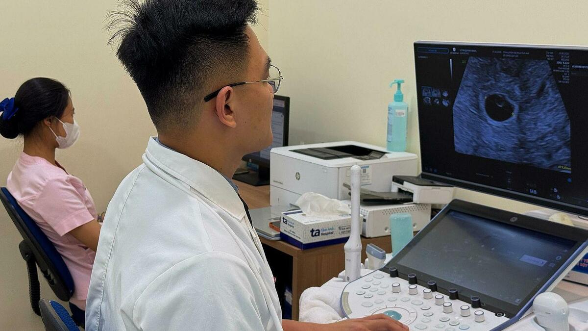
Doctor Loi is doing a 2D ultrasound for a pregnant woman. Illustration photo: Tam Anh General Hospital
4D ultrasound recreates three-dimensional images in real time, allowing a clearer view of the face, limbs, and movements of the fetus in the womb. Visual images help support the assessment of some morphological abnormalities such as cleft lip, cleft palate, abnormalities in the limbs or abdominal wall.
However, 4D ultrasound does not replace 2D ultrasound. In obstetric practice, doctors still rely mainly on surveys and indicators on 2D ultrasound to assess fetal health. 4D ultrasound is a supporting tool to help observe morphology more intuitively at necessary times.
Pregnant women are often recommended to have 4D ultrasound performed at three important milestones in pregnancy.
From 11 weeks to 13 weeks and 6 days: This is the time when the doctor measures nuchal translucency, determines gestational age, evaluates the risk of chromosomal abnormalities, and initially surveys the formation of fetal organs.
From 18 to 22 weeks (also known as the fetal morphology ultrasound milestone): This is an important stage to examine in detail the brain, heart, spine, face, abdominal wall, kidneys, limbs and many other organs, thereby detecting many birth defects early, if any.
From 28 to 32 weeks: The doctor evaluates the development of the fetus, the position of the placenta, the amount of amniotic fluid, the umbilical cord, and monitors for abnormalities that may appear or become more evident in the later stages of pregnancy. At this time, images of the fetus’s face and movements often appear more clearly.
After this milestone and in the last weeks of pregnancy, pregnant women often have regular ultrasounds using 2D ultrasound to monitor fetal weight, fetal position, placental position, amount of amniotic fluid and factors related to the birth process. The doctor can evaluate the condition of the fetus, predict the possibility of a normal birth or prescribe appropriate intervention methods when necessary.
The important thing for pregnant women is not to choose 2D or 4D ultrasound, but to have a full prenatal examination and ultrasound as prescribed by the doctor. Early detection of fetal abnormalities depends more on the timing of the ultrasound, the level of the doctor performing it, and the quality of the equipment than on the type of ultrasound alone.